About Field of View
Having the most specific information on your patient’s condition is vital to proper treatment. With a cone beam computed tomography (CBCT) scan, you can get a more detailed view of the mouth including precise tooth position, bone dimension and quality, TMJ disorders, and other dental conditions. Field of view (FOV) is the anatomical area that is included in the 3D data volume. This is the area where the patient will receive radiation.
Classifications for the size range include small, medium, and large. The ALARA Principles (As Low As Reasonably Achievable) govern the best use of radiation for patients.
Area Captured by Small Field of View Cone Beam
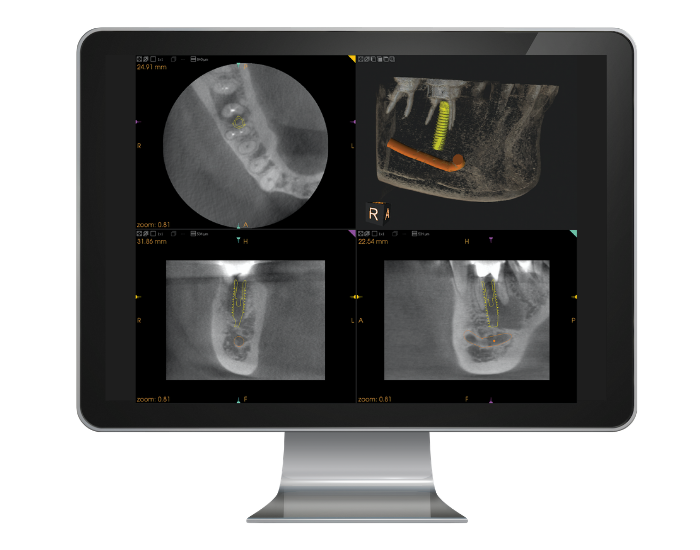
Small field of view cone beam systems capture an area of approximately 2-5 teeth and their surrounding anatomical structures. This results in less volume to interpret as well as minimized radiation dose for the patient. Small FOV cone beam is typically adequate for 3D periapical evaluations of the selected teeth, alveolar bone, and a limited amount of maxillary or mandibular basal bone.
Indications for Small Field of View Cone Beam
Endodontics are the most common use for small FOV scans because they offer high spatial resolution and give the practitioner the ability to see changes in the periodontal ligament spaces or lamina dura, root fractures, periapical lesions, the relationship of an impacted tooth to its surrounding anatomical structures, and root canal morphology.
Because it has such a focused area, small FOV CBCT is also the ideal choice for placing single implants or completing surgical procedures.
Small FOV CBCT systems are often a good entry point into 3D imaging due to their lower cost over larger FOV options. In fact, we’ve seen practices start with a small FOV unit and then upgrade to a larger system later. Renew Digital’s low initial price point as well as our trade-in options make this a favorable option for many practices that are new to cone beam imaging.
Limitations of Small Field of View Cone Beam
Some small FOV cone beam options have the ability to create a larger volume that includes the complete arch. This eliminates the need to capture several smaller images and stitch them together. In the stitching process, multiple adjacent focused field of view scan are “stitched” together in reconstruction to create one dental arch.
The main disadvantage of this technique is the amount of patient exposure to radiation during multiple CBCT scans. Another disadvantage is the potential for error or miscalculation during the stitching process. With a single scan of the entire area, you can eliminate room for error and reduce patient exposure.
Get Started Today!
Whether this is your first cone beam unit or your fifth, Renew Digital is here to help you find the perfect small field of view cone beam option for your practice and budget. Our Sales Team are experts at understanding the clinical needs of our customers and finding the right fit for you, both now and in the future. Plus, they can help save you up to 30-50% off the price of a new small FOV cone beam system.
Every purchase includes expert installation and training, warranty, and customer service. Please contact us today to learn more about our current availability and choose your small field of view cone beam unit.
